On the hunt for what you can’t see in the Connecticut River
By Sally Warring
Scientific images by Sally Warring
Additional photography by Jody Dole
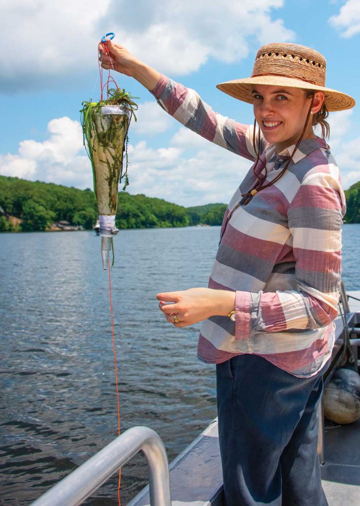 On a hot weekend in July, we headed out on the Connecticut River for a wildlife safari. We weren’t aiming to see osprey, swallows, bald eagles, or turtles, though we saw plenty of those. Instead, we were on the hunt for the River’s smallest inhabitants… its microbial wildlife. The presence of such wildlife can be an indicator of the health of a river.
For a microbial safari one needs only a receptacle to hold a water sample, a pipette for transporting a small amount of the water sample onto a glass microscope slide, a glass coverslip to hold it in place and, of course, a microscope.
Microbial wildlife thrive all around us. These organisms are tiny, single-celled creatures that lead complex lives and play important roles in our ecosystems. Most are no more than one tenth of a millimeter long, well below the threshold for visibility by the human eye. With a microscope, we can magnify these creatures one hundred, two hundred, or even four hundred times to observe their forms and behavior.
When hunting for microbes there are some signs visible to the naked eye: when microbes grow to large populations, their “communities” can become visible. On our River trip, we were on the lookout for signs of such microbial communities.
Color is an important sign. Microbes come in greens, browns, reds, yellows, and whites, and when you see water or a surface that has taken on one of these hues, chances are you’re looking at a microbial ecosystem.
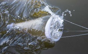 Once on the River, we scooped up some water that was covered in a whitish foam and streaked with brown. Foam is another sign of microscopic lifeforms. Foam is produced when the movement of the water churns up organic matter, mostly proteins and fats, from suspended microbial organisms. Sure enough, once we got this foamy water under the microscope, we found the cause of these visual aquatic phenomena: Diatoms.
Diatoms are beautiful single-celled microbial algae. They are common in freshwater, saltwater, and everywhere in between. All algae diatoms live off sunlight. Inside each cell is a golden-brown chloroplast, the same ingredient that converts sunlight, water, and carbon dioxide into food for plants. The diatoms float in the upper strata of water where light is plentiful, making sugars from light energy. This produces much needed oxygen; world-wide it is estimated that diatoms provide 20 percent of global oxygen.
Diatoms are expert builders. Each diatom absorbs silica from the environment, then extrudes that silica to create a shell around its soft single-cell body. These glass shells are called frustules. Many are intricately shaped and beautifully decorated. Some shapes help with flotation and movement, while other features allow for the exchange of gasses and material between the cell and its surroundings.
Some diatoms move by excreting a kind of mucus out of a groove called a raphe; the cell uses this mucus to slide over surfaces. You can find surface-dwelling diatoms all through the River. The next time you see a submerged plant, look to see if it’s coated with a brown or soil-like layer; that layer is probably from diatoms that have attached to the submerged plant.
Diatoms sit at the base of the food chain providing meals for other microbes and zooplankton. When a diatom dies or gets eaten, the tiny silica frustule remains and falls to the sediment layer, slowly building up over time. Under the right conditions that sediment layer can harden and fossilize into a useful sedimentary rock known as diatomite or diatomaceous earth.
In the pictures here, the diatoms with golden-brown colors are living; the ghostly clear shapes are the empty frustules. Each differently-shaped diatom or frustule represents a different species of diatom. There are around 12,000 diatom species currently described and probably many more to discover. As we observed, many diatom species call the Connecticut River home.
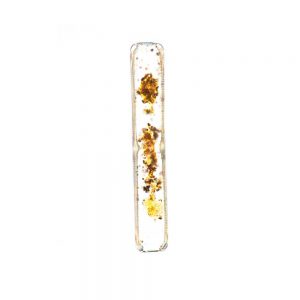
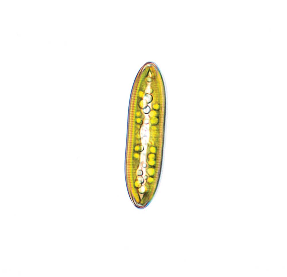
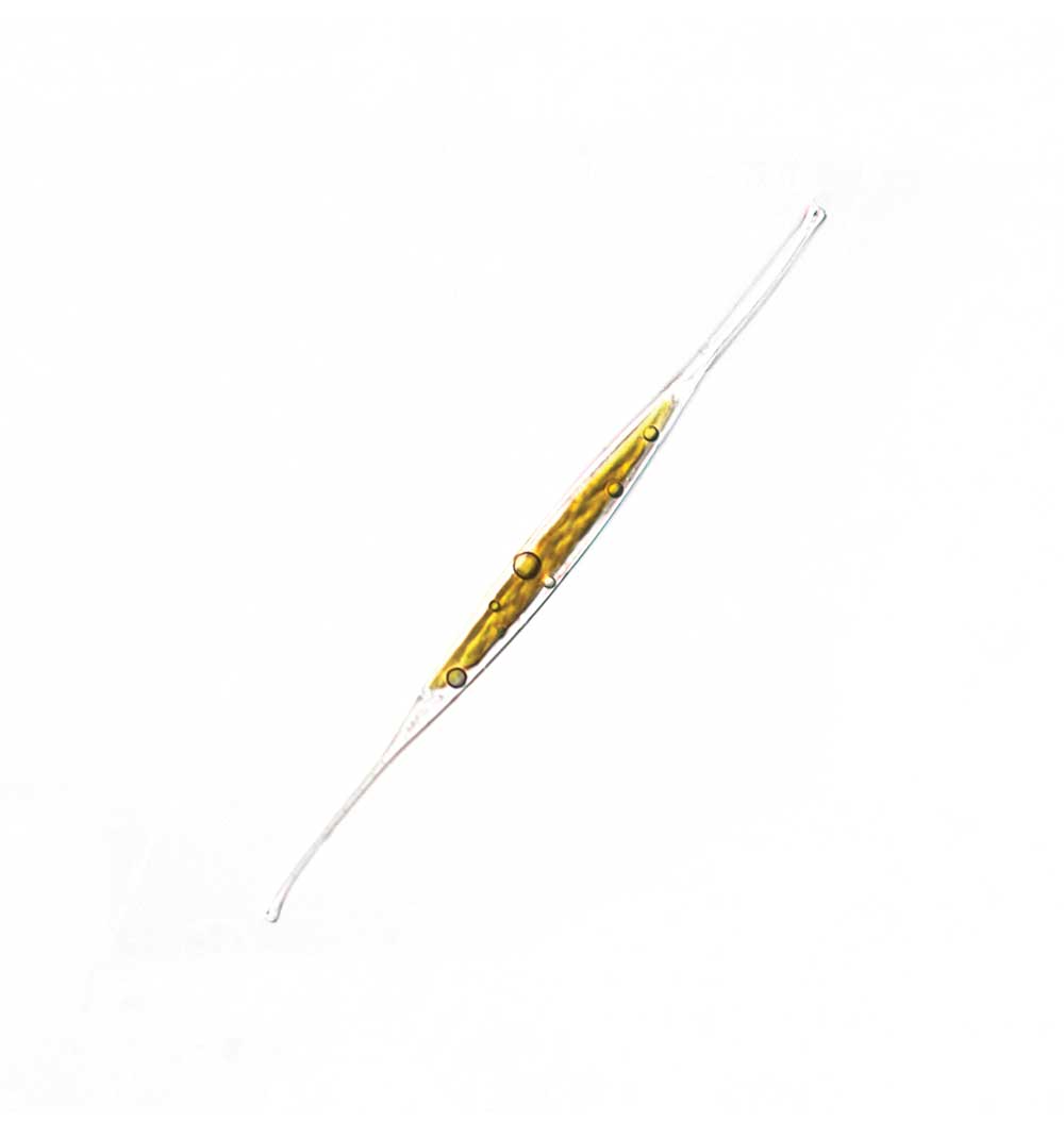
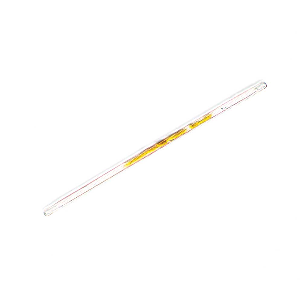
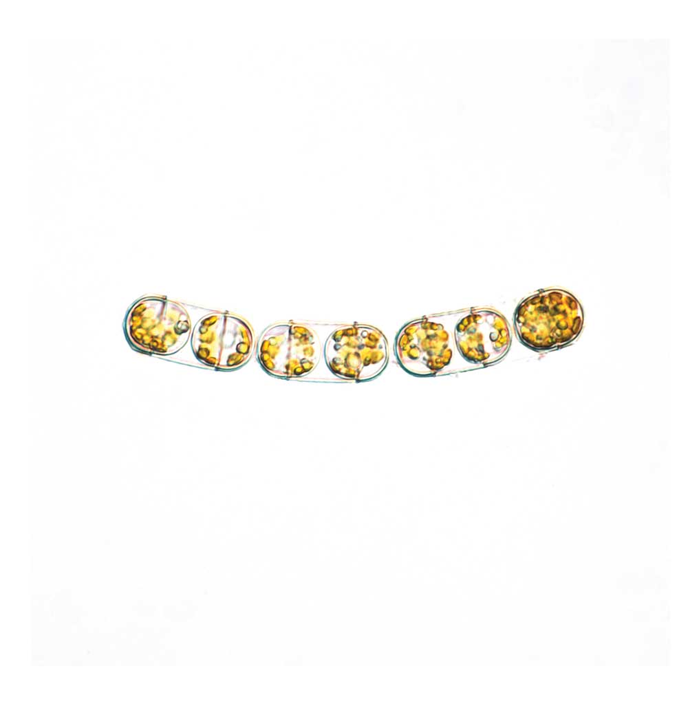
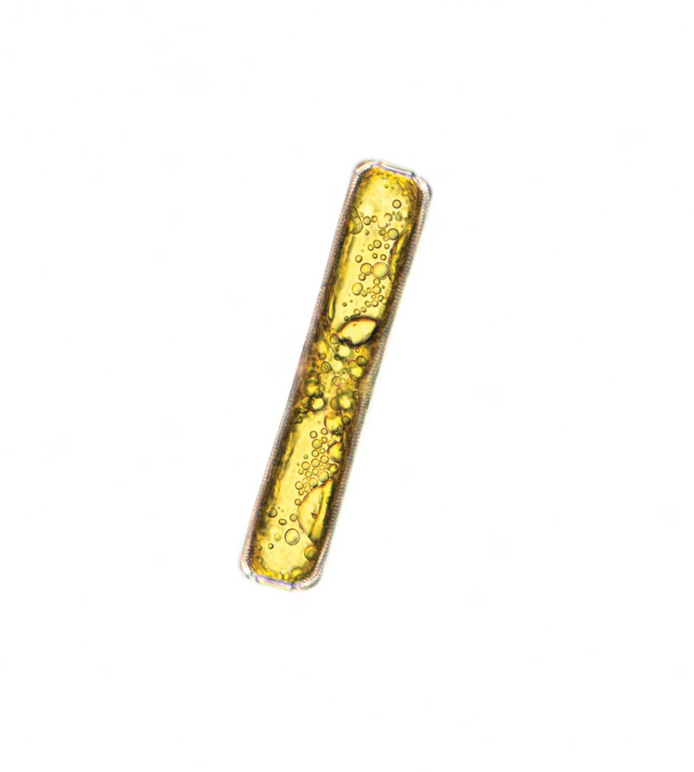
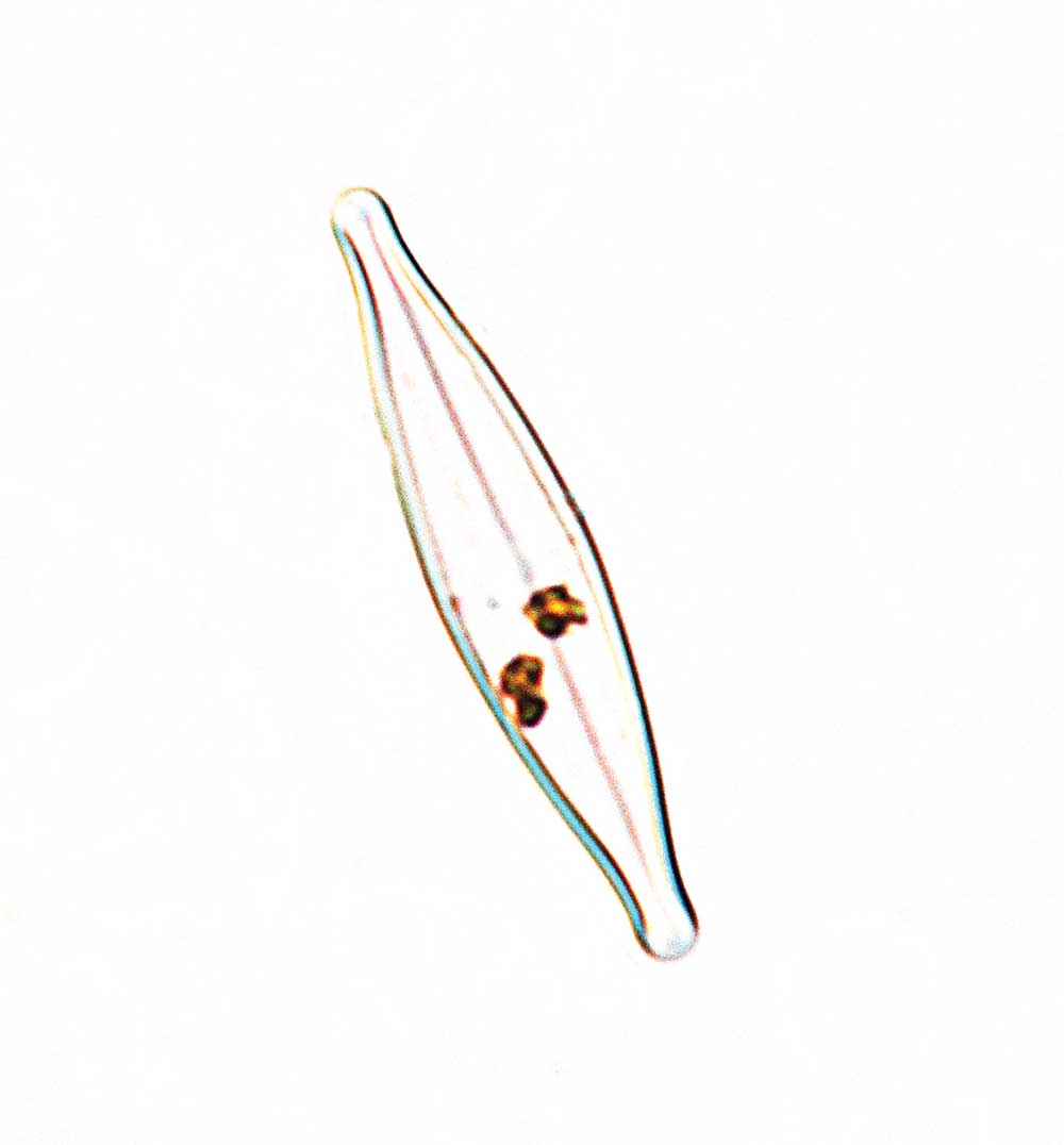
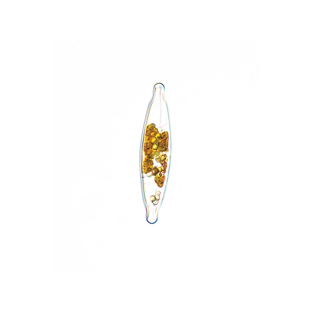
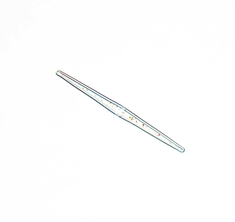
Diatoms are beautiful single-celled microbial algae